
MIT neuroscientists have created a way to measure dopamine production in the brain for over a year. The new method could help doctors learn more about dopamine’s role in brain function.
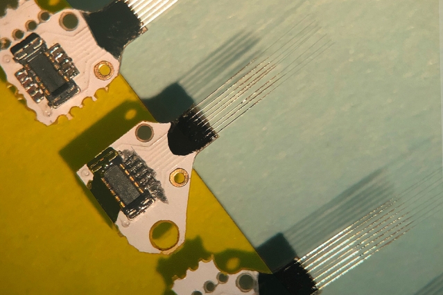 MIT neuroscientists have devised a way to measure dopamine in the brain for more than a year, using microfabricated sensors that are so tiny they don’t lead to the formation of scar tissue. Source: Felice Frankel
"Despite all that is known about dopamine as a crucial signaling molecule in the brain, implicated in neurologic and neuropsychiatric conditions as well as our ability to learn, it has been impossible to monitor changes in the online release of dopamine over time periods long enough to relate these to clinical conditions," says Ann Graybiel, an MIT Institute Professor, a member of MIT's McGovern Institute for Brain Research and one of the senior authors of the study.
Traditional dopamine measuring systems consist of carbon electrodes that have a shaft diameter of around 100 microns. While this seems small, it is too big to be used long-term. These systems can only be used for about a day because they create scar tissue that interferes with the electrode’s operation.
In 2015, MIT’s team proved that microfabricated sensors could be used to measure dopamine directly in the striatum in the brain, where dopamine is produced. The striatum has dopamine-produced cells that are vital in habit formation and reward-enforced learning.
The newly developed, microfabricated probes measure at around 10 microns in diameter. Because they are so small, the team could implant 16 sensors at a time without developing scar tissue. The sensors measure dopamine production in different areas of the striatum. The new study found that this method could be used for years at a time without the body interfering with operations.
"Our fundamental goal from the very beginning was to make the sensors work over a long period of time and produce accurate readings from day to day," said MIT postdoc and lead author Helen Schwerdt. "This is necessary if you want to understand how these signals mediate specific diseases or conditions."
During the study, the researchers implanted three to five sensors per animal they were testing. The sensors were placed 5 mm deep in the striatum. The team took readings every few weeks. They found that the sensor's measurement stayed consistent for up to 393 days.
"This is the first time that anyone's shown that these sensors work for more than a few months. That gives us a lot of confidence that these kinds of sensors might be feasible for human use someday," Schwerdt says.
These sensors could be used to monitor Parkinson’s disease patients who receive deep brain simulations. Deep brain simulations are implanted electrodes that deliver electrical impulses deep in the brain to combat the effects of Parkinson’s. Sensor monitoring of dopamine could help doctors deliver direct simulations in selective areas of the brain that are being affected.
The next step is to adapt the sensors to measure other neurotransmitters and measure the electrical signals in the brain.
"Understanding those relationships between chemical and electrical activity will be really important to understanding all of the issues that you see in Parkinson's," Schwerdt says.
The paper on the new sensor research was published in Communications Biology.
