When it comes time for the U.S. Food and Drug Administration (FDA) to test new drugs, the process typically takes many years and billions of dollars. A team of engineers from University of California, San Diego has 3D-printed a tissue that closely mimics the human liver that could potentially be used for patient-specific drug screening and disease modeling, to combat the process.
According to the researchers, the advance could help pharmaceutical companies save time and money when developing new drugs. "We've made a tool that pharmaceutical companies could use to do pilot studies on their new drugs, and they won't have to wait until animal or human trials to test a drug's safety and efficacy on patients. This would let them focus on the most promising drug candidates earlier on in the process,” said Shaochen Chen, Nano Engineering professor at the UC San Diego Jacobs School of Engineering.
Other groups have been working on liver models, as well, since the liver plays a vital role in how the body metabolizes drugs. To date, no other model has consisted of the complex micro-architecture and diverse cell makeup of a real liver.
How they did it
The UC San Diego team, led by Chen and Shu Chien, a professor of Medicine and Bioengineering, employed a novel bio-printing technology capable of rapidly producing complex 3D microstructures that mimic the features of biological tissues.
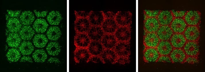 Images of the 3-D-printed parts of the biomimetic liver tissue. (Image Credit: Chen Laboratory, UC San Diego)
To print the liver tissue, the team first printed a honeycomb pattern of 900-micrometer-sized hexagons, each containing liver cells derived from human induced pluripotent stem cells. Then it printed endothelial and mesenchymal supporting cells in the spaces between the stem-cell-containing hexagons.
The entire structure, with dimensions of 3 × 3 millimeter square and 200 micrometers thick, took only seconds to print, which the team considers “a vast improvement over other methods to print liver models, which typically take hours.”
The researchers then tested the resulting tissue's ability to perform different liver functions, such as albumin secretion and urea production. When compared to other models, they found that their model was able to maintain these functions over a longer time period than other liver models.
The 3D printing technique employed here may also be found useful when it comes to studying other diseases that affect the liver. "The liver tissue constructed by this novel 3D printing technology will also be extremely useful in reproducing in vitro disease models such as hepatitis, cirrhosis and cancer," said Chien. "Such realistic models will be invaluable for the study of the pathophysiology and metabolic abnormalities in these diseases and the efficacy of drug therapies."
