The application of an unconventional imaging technique to the spectroscopic measurement of a gas molecule shows promise for the measurement of methane and other greenhouse gases.
Researchers coupled the ghost imaging method with a supercontinuum light source to capture the wavelength-dependent light transmitted through samples, and demonstrate that the technique can measure the spectral signature of methane with subnanometer resolution.
Ghost imaging produces images by correlating the intensity of two light beams that, taken individually, do 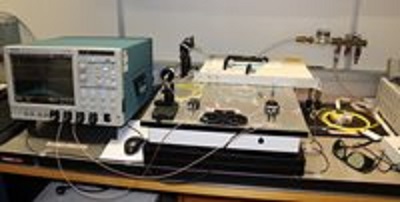 The new ghost imaging approach uses a pulsed supercontinuum light source (right), with large spectral fluctuations. An oscilloscope (left) was used to measure the reference spectral fluctuations and the total signal transmitted through the gas. Source: Caroline Amiot, Tampere University of Technologynot deliver meaningful information on the shape of the object, but instead allow indirect inferences about its properties. Some distortions associated with typical imaging systems in harsh environments can be eliminated, and the process is useful for creating high-resolution images of physical objects.
Gas molecules are often sparse and only change the total light transmittance by a small amount, pointing to the need for powerful light sources or extremely sensitive equipment to detect them. Ghost imaging enables measurement with less powerful light sources since it detects an integrated signal containing many wavelengths, in contrast to one wavelength typical of traditional spectroscopy methods.
The spectral image generated captures the transmission or reflection spectrum of an object by correlating two arms of a beam of light -- one that encodes a random pattern that acts as a probing reference and the other that illuminates the sample. The new approach uses a supercontinuum light source, which emits pulses that each contain many wavelengths of light. The random fluctuations that occur between the spectra associated with consecutive pulses are used to create the reference necessary for performing spectral ghost imaging.
The light transmitted through a sample is then sensed with a fast detector without spectral resolution that provides an integrated signal for all the wavelengths of the spectral bandwidth under consideration. The image initially appears as a noisy blob, but once it is correlated with the reference spectral fluctuations, the spectral image begins to appear.
The technique was applied to the production of a spectral image of methane. The researchers observed that the ghost imaging measurements perfectly reproduced the series of discrete absorption lines that are the fingerprints of methane, and matched well with more conventional direct spectroscopy measurements performed for comparison.
Efforts now focus on controlling the spectral fluctuations using pre-programmable light sources that would remove the need to measure the reference spectral patterns. The use of spectral-domain ghost imaging with an optical coherence tomography setup is also being pursued to allow sensitive information to be gained from tissue or other biological samples without using damaging amounts of light.
Scientists from Tampere University of Technology in Finland, the University of Eastern Finland and the University of Burgundy Franche-Comté in France participated in this research, which is published in Optics Letters.