A team from Privolzhskiy Research Medical University has further developed a fluorescence imaging technique to take it from limiting, tiny millimeter-sized samples to less restricted macro-sized samples. The system was developed at Becker & Hickl GmbH, and biological testing was done at Privolzhskiy Research Medical University. The new approach expands the sample sizes from a millimeter to around four square centimeters. The technique could be used in clinics as a sensitive and precise method to identify tumor edges during surgery. The new imaging approach is based on fluorescence lifetime imaging microscopy (FLIM).
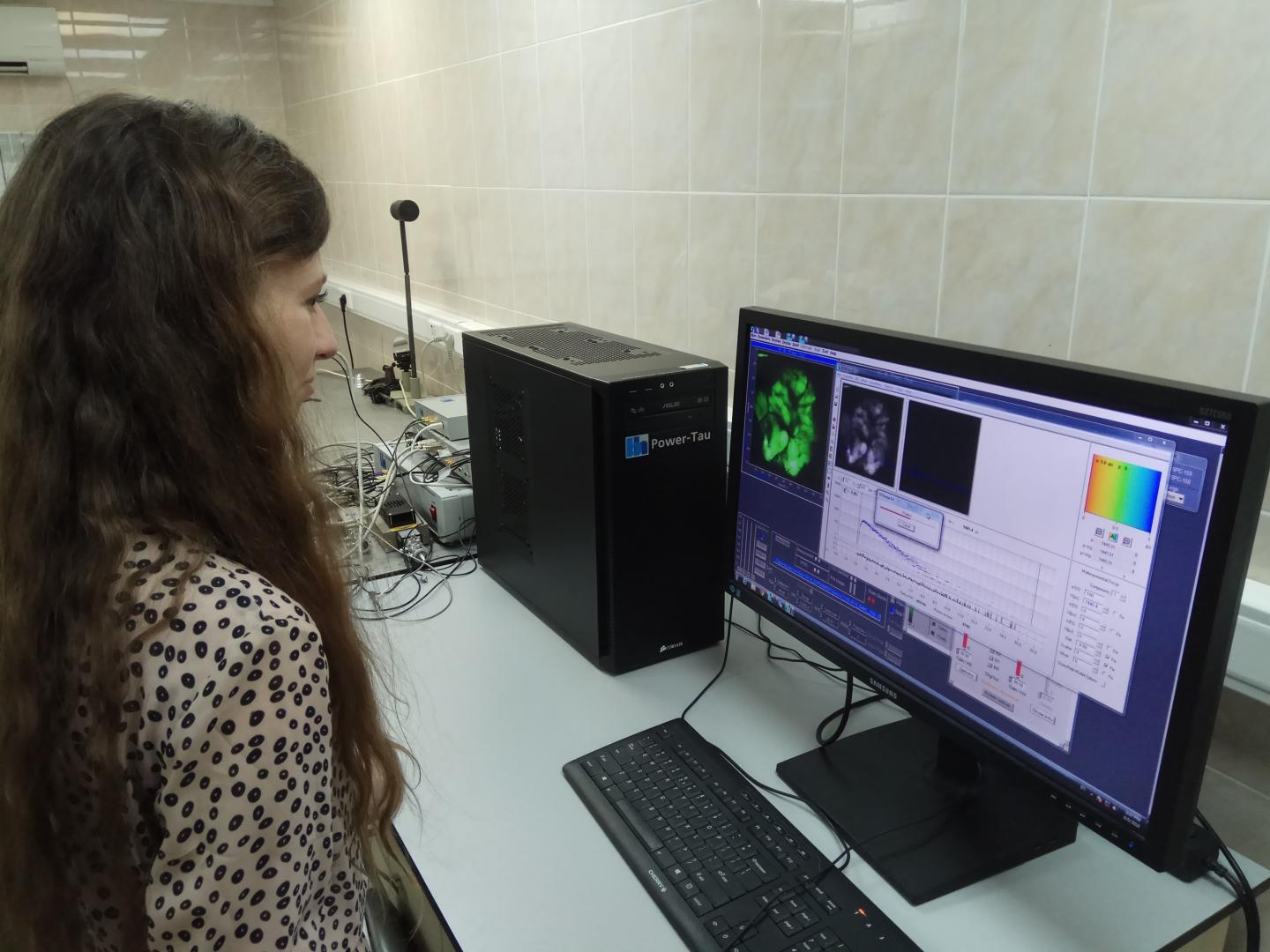 Researchers have developed a macro-FLIM system that can analyze samples with areas up to 4 square centimeters. Source: Vladislav Shcheslavskiy
"Our macro-FLIM system can not only obtain a sample's structural information but also allows observation of certain biochemical processes taking place within the sample," said Becker & Hickl senior research scientist Vladislav Shcheslavskiy. "Although our goal is to develop this for clinical use, it could also be very useful in fundamental studies for probing biological processes as the disease develops or investigating biological responses to different types of therapy."
The system has been used to observe metabolic processes in a live tumor which was completely impossible with the previous systems. The FILM system can be used to analyze other samples with large areas that have been previously unable to be analyzed with the microsystem. The non-destructive method could even be used with paintings that need to be restored in order to find out what is underneath.
FLIM precisely measures the fluorescence decay rate of naturally fluorescent molecules, tags or labels that are added to a tissue, like a tumor. FLIM is used to gather information about the properties of a molecule and micro-environments.
FLIM is normally performed with a laser scanning confocal microscopy system to created FLIM images at the macroscale. The system scans a laser beam across a fluorescent sample and creates an image. The system incorporates lasers with picoseconds long laser pulses that are incredibly sensitive.
"Careful optical design along with the picosecond lasers, sensitive and fast detectors and fast single photon counting electronics allowed us to record fluorescence decay with high precision at the macroscale,” said Shcheslavskiy.
Confocal microscopy has been limited to a millimeter imaging area until now. The researchers have plotted photon distributions across a large area of the sample for the first time. The system has been used to make images of fluorescent microbeads that have a diameter of 14.6 microns.
The researchers have used the FLIM system to analyze an entire tumor within a live mouse. This measures the fluorescent lifetime of the genetically encoded red fluorescent protein. The team was able to identify the tumor and nicotinamide adenine dinucleotide (NADH) with the system.
"The sensitivity of our system was high enough to observe fluorescence of intrinsic tissue components such as NADH without any labeling," said Shcheslavskiy. "In addition to being used to study metabolism in a tumor, macro-FLIM could be used to follow cell death or oxygen status of tumors on a macro scale with cellular resolution."
The next step for the team is to further develop the system to work for clinical applications. The team says the system needs to improve flexibility and mobility before it can work in doctors' offices or labs. The researchers also want to combine the macro-FILM system with the scanning stage so FILM can be performed in areas that are 10x10 centimeters long.
The paper on the new system was published in Optics Letters.
