 The augmented reality system in use. Source: Imperial College London Last week, we reported on a group of researchers in Canada that were using augmented reality that moves with a patient through motion tracking and infrared cameras to help doctors see what is happening inside the body without invasive processes.
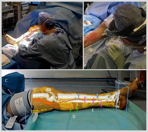
Now, scientists at the Imperial College London at St. Mary’s Hospital are using augmented reality through a Microsoft HoloLens headsets while operating on patients undergoing reconstructive lower limb surgery. The team used the technology to overlay images of CT scans onto each patient’s leg allowing surgeons to see through the limb during surgery.
Through trials, researchers were able to use the approach to help surgeons locate and reconnect key blood vessels during reconstructive surgery, which they believe could improve outcomes for patients.
Following a traumatic event, patients may have tissue damage or open wounds that require reconstructive surgery using fasciocutaneous flaps. These flaps of tissue, which are taken from elsewhere on the body and include skin and blood vessels, are used to cover the wound and enable it to close and heal properly.
The standard approach to doing this is to use a handheld scanner which uses ultrasound to identify blood vessels under the skin by detecting the movement of blood pulsing through them. Augmented reality offers a new way to find these blood vessels quickly and more accurately by overlaying scan images onto the patient during the operation, researchers say.
"We are one of the first groups in the world to use the HoloLens successfully in the operating theatre," says Dr Philip Pratt, a Research Fellow in the Department of Surgery & Cancer. "Through this initial series of patient cases we have shown that the technology is practical, and that it can provide a benefit to the surgical team. With the HoloLens, you look at the leg and essentially see inside of it. You see the bones, the course of the blood vessels, and can identify exactly where the targets are located."
How It Works
During the trials for the technology, five patients needing reconstructive surgery on their legs had CT scans to map the structure of the limb. Images from the scans were then segmented into bone, muscle, fatty tissue and blood vessels and loaded into intermediary software to create 3D models of the leg,
These models were then put into software that renders the images for the HoloLens headset, which then overlays the model onto what the surgeon can see during surgery. During the operation, staff can manipulate these augmented reality images through hand gestures to make any fine adjustments and correctly line up the model with surgical landmarks on the patient’s limbs such as the knee joint or ankle bone.
"Now, using the HoloLens, we can identify where the blood vessels are in 3D space and use virtual 3D arrows to guide the surgeon,” says Dr Dimitri Amiras, a consultant radiologist at Imperial College Healthcare NHS Trust. “Currently, data preparation is a time consuming process, but in the future much of this could be automated, with the consultant radiologist checking the accuracy of the model against the original scan.”
The full research can be found in the journal European Radiology Experimental.
