 Principle of the PBM technique. Source: Xiang Zhang
Nanoscale imaging is important to many modern applications in materials science, physics, biology, medicine and other fields. But the current techniques are limited because of their resolution, imaging speed or the inability to look behind opaque objects with subjective shapes.
This imaging would be useful for many applications like investigating spongy electrodes, helping increase capacity and charging speed of the next generation of batteries.
The team behind this research was led by professor Xiang Zhang from the University of California in Berkeley. They demonstrated a method of stunning properties.
"We wanted to overcome limitations of current nano-imaging techniques and are excited to have found a way to image complex 3D nanostructures even with intricate internal structures such as cavities," explains Zhang.
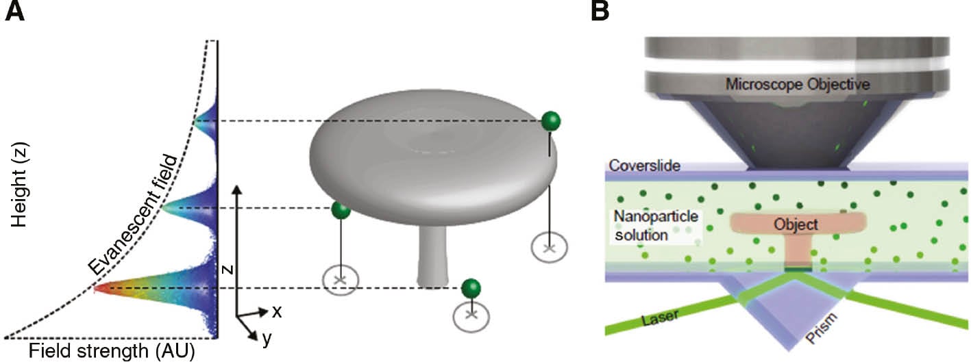
Nanoparticles are covered in a fluid that surrounds the object that is being investigated. By exploiting the special properties when interacting with light, each particle acts as a light source, and proves the object from all sides, behind overhangs and inside any cavities. It has a resolution of 30nm in all directions. The new technique offers true 3D imaging at the nanoscale.
Other than the applications in the technology sector, plasmonic Brownian microscopy might be used to map out the biological machinery inside single cells, especially those with intricate interval structures. This would help further the understanding of the basic mechanisms of living organisms and might be key to a rise in new medical solutions.
A paper on this research was published in Nanophotonics.
