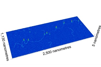
 A typical 3D image collected by the nanomapping microscope showing a CRISPR-labelled DNA molecule. Image credit: University of BristolA new nanomapping microscopy technique developed by an international team of researchers could transform the way disease-causing genetic mutations are diagnosed and detected.
The microscope maps hundreds of chemically barcoded DNA molecules every second. CRISPR-Cas9 is used to label the molecules so that they can be mapped almost as accurately as DNA sequencing, while also processing large sections of the genome at a much faster rate. An atomic force microscope was modified with off-the-shelf DVD components so that it could physically map the lengths of individual DNA molecules to a resolution of tens of base pairs at rates of hundreds per second. This speed increase enables this DNA barcoding method to be used for real-world diagnostics for the first time.
The system uses a microscopic stylus—similar to a needle on a record player— that barely makes contact with the surface of the material being studied. The interaction between the stylus and the molecules generates the image as the microscope measures single DNA molecules with sub-atomic resolution. The chemical barcoding method alters the chemical reaction conditions of the CRISPR enzyme so that it only sticks to the DNA and does not actually cut it.
"Using the laser focussing mechanism found in every DVD player we have built a microscope that has the resolution and speed to measure every molecule on the sample surface in 3D," said Dr. Oliver Payton from the University of Bristol's School of Physics.
"Although other types of microscope have the resolution to see these DNA molecules they are thousands of times slower and it would take years to make a confident diagnosis. Not only is our microscope perfect for these medical applications, but because of the readily-available DVD player components it can be mass-produced."
The method was demonstrated by mapping genetic translocations present in lymph node biopsies of lymphoma patients. Such translocations are especially prevalent in blood cancers such as lymphoma but occur in other cancers as well.
The University of Bristol team is also using the microscope to solve a diverse range of nanoscale challenges across fields such as 2D materials, corrosion and life sciences. It is also being commercialized via the University of Bristol spin-out, Bristol Nano Dynamics Limited.
Scientists from Virginia Commonwealth University, New York University and the University of California-Los Angeles also participated in this research, which is published in Nature Communications.
