A simple tweak to the standard light microscope yields a smart instrument that can adjust its settings and improve the speed and accuracy of image-based medical diagnoses. A new illumination scheme augmented by a deep neural network enables the microscope to optimize its own lighting conditions for the type of sample to be analyzed.
A programmable LED array added to the microscope is controlled by a convolutional neural network, which 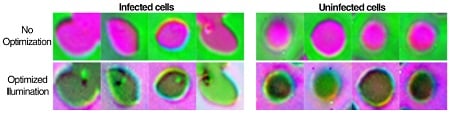 Compared to a traditional microscope (top), the red blood cell images created by the new microscope (bottom) contain more noise, but the malaria parasites are lit up by bright patches due to optimized lighting conditions. Source: Duke Universitydetects the presence of infection in samples and produces the ideal LED illumination pattern to highlight sample features essential for classification. The approach was tested with hundreds of samples of malaria-infected red blood cells prepared as thin smears, in which the cell bodies remain whole and are spread out in a single layer on a microscope slide. The machine learning algorithm keyed in on highlighting features important for diagnosing the disease.
A specific ring-shaped LED pattern of different colors emitted from relatively high angles generated microscope images that correctly classified the malaria parasite with a 90% accuracy, exceeding the 75% accuracy posted for trained physicians and other machine learning algorithms.
The technology described in Biomedical Optics Express can be applied to other diagnostic imaging tasks, potentially automating entire processes performed in hospital pathology labs. Researchers from Friedrich-Alexander University (Germany), Duke University, Y Combinator Research, Humboldt University (Germany) and Fraunhofer IISB (Germany) contributed to this development.
