Computed tomography (CT) scans are used to reconstruct a 3D organ from multiple 2D image slices for diagnostic assessments. Efforts to translate this technology to the molecular level could further precision medicine and the ability to customize treatments to each patient’s unique cellular features.
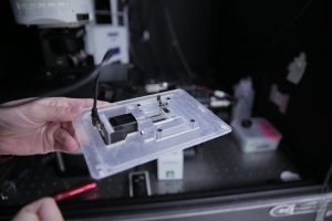 A 3D-printed custom device holds a motor (left) that spins the tube containing the sample. Source: Renee MeillerProgress in this challenging task is reported by University of Wisconsin-Madison researchers, who have succeeded in the 3D imaging of collagen, a protein essential for connective tissue and bone stability.
The challenge in imaging collagen is that these molecules are transparent. The contrast between lighter and darker objects that a traditional optical microscope reads won’t be generated by collagen. Second harmonic generation microscopy can convert the signals associated with collagen into 2D images, but not 3D.
The researchers developed an experimental and computational framework for assembling 2D collagen images, taken from multiple angles around the tissue sample, into a moderate-resolution 3D view, similar to the familiar CT scan of human organs. A 3D-printed device holds a tube attached to a small motor and sits on the stage of an upright microscope. Once a tissue sample is placed into the tube, the motor starts to spin it. Every time a laser source, located below the stage, sends light through the rotating sample, a laser scanner records the resulting 2D microscope image. At the end of the procedure, a complex mathematical algorithm reconstructs a 3D image from all of the 2D slices.
This technology might be used to detect subtle differences between highly aligned collagen fibers in breast and ovarian cancer tissue, which are distinct from the cross-hatched mesh of collagen found in normal tissue. These images may inform treatment decisions not only for cancer, but also for pulmonary fibrosis.
