A collaboration between CEA-Leti and Siemens Healthineers has resulted in the development of a new X-ray photon-counting detector module (PCDM). When integrated into an X-ray computed tomography (CT) scanner prototype from Siemens Healthineers, the module was demonstrated to deliver improved spatial resolution, decreased radiation or iodine contrast dose requirements and reduced levels of image noise and artifacts.
CEA-Leti used its simulation tools to design the geometry of the PCDM and select a semiconductor based on cadmium telluride (CdTe). Current X-ray CT scanners produce images with energy-integrating detectors, which are based on indirect conversion technology. PCDMs instead directly convert X-ray photons into electronic 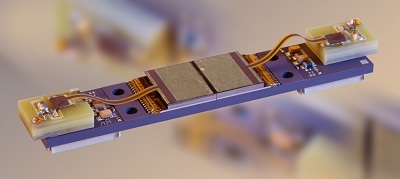 The X-ray photon-counting detector module. Source: CEA-Letisignals with a higher conversion yield.
The use of CdTe supports simultaneous acquisition of high-spatial-resolution and multi-energy images. Higher spatial resolution improves image quality by using a small-pixel size detector and multi-energy provides color images, compared to grey-level images of conventional detectors, and allows a precise determination of the atomic number of any chemical elements present in the body. Clearer images of very-fine structures, such as small airways in the lungs and thin wires in coronary stents, are obtained.
Evaluations conducted at the Mayo Clinic confirm improved spatial resolution, lower radiation or iodine contrast dose requirements and decreased levels of image noise and artifacts. Researchers demonstrated the simultaneous acquisition of multiple 150-micron-resolution datasets, each representing a different energy spectrum.
