An international team of experts from 26 institutions around the world has further developed cell sorting techniques. The technique, called image-activated cell sorting (IACS), uses optical, microfluidic, electrical, computational and mechanical technology. IACS sorts cells based on their global phenotypic profiles and spatial and morphological properties.
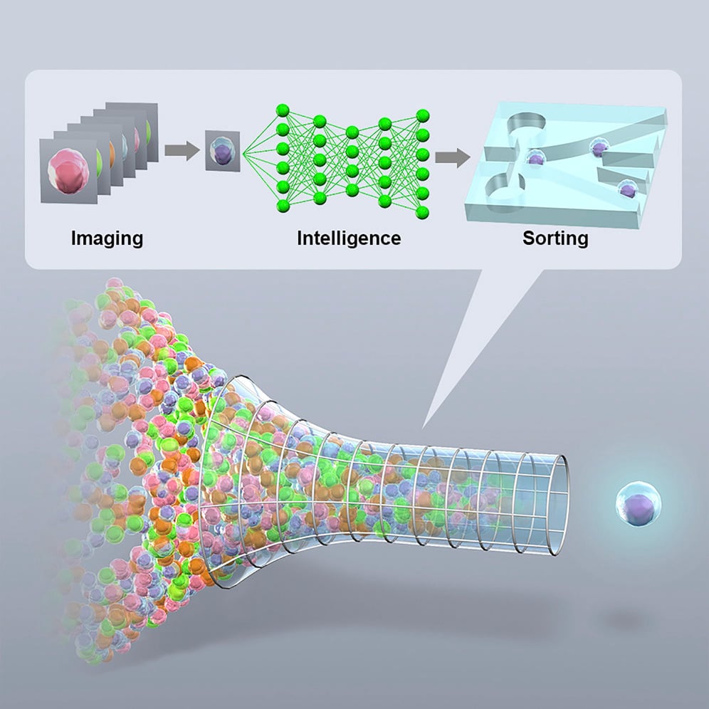 Image of how the cell sorting technique works. Source: University of Tokyo
"We aim to extend flow cytometry's capability from 1D intensities to 2D pictures to sort cells with unique spatial architectures of biomolecules. This will allow addressing new fundamental biological questions like 'How is cellular architecture molecularly connected with physiological function?'" says senior author Keisuke Goda, a physical chemist at the University of Tokyo. "We envision the developed tool to be broadly applicable in the study of what genes affect the spatial localization of various molecules within cells."
IACS requires a perfect balance between speed and accuracy. The new method isolates target cells in real-time without interruptions by using deep learning to process the data. IACS took two years to design, two years to develop the subsystems and two years to integrate and test. IACS was tested using microalgae and blood cell samples.
During the cell sorting process, a tube with suspended cells is put at the injection point of the IACS system. The cells then go through a cycle of the system, where they are imaged one by one under a microscope. The data is collected and informs the sorting decision making. The system then decides if the cells meet the given criteria; if they do they are separated from the rest of the cells. At the end of the cycle there are two tubes: one filled with sorted cells and the other filled with unsorted cells. These tubes are inspected under an optical microscope and evaluated for quality.
"The platform enables image acquisition, image processing, decision making, and actuation, all within 32 milliseconds even with deep learning algorithms, and hence realizes real-time image-based intelligent cell search and sorting at an unprecedented rate of about 100 cells per second," Goda says. "The intelligent IACS technology is highly versatile, can handle various types and sizes of cells in diverse fields ranging from microbiology to hematology, and holds promise for making machine-based discoveries in biological, pharmaceutical, and medical sciences."
The IACS platform can currently only analyze individual cells. The system can’t handle larger biological objects, but the researchers plan to modify the system to allow for larger analyzation samples.
Along with the development of IACS, the team is launching an open innovation platform. The sorting machine is huge and difficult to assemble, so it is difficult to use outside the lab. This platform allows other researchers and developers to submit their ideas and samples to be tested with IACS without having to ship the machine or send a scientist out to the University of Tokyo in Japan.
The study on the new system was published the journal Cell.
