Congenital heart disease (CHD) strikes approximately nine in every 1,000 live births. Overall survival rates have steadily increased, yet patients with CHD continue to face many health obstacles and challenges — including the risk of long-term morbidity, the need for re-intervention and hospitalization, neurodevelopmental outcomes and more.
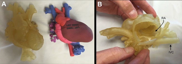
One emerging technology that impacts the way cardiologists treat their CHD patients is 3D printing. The technology enables a 3D replica of a patient's anatomy for precise pre-surgical planning and simulation, which can potentially reduce time spent in the operating room and result in fewer complications.
According to a recent review paper, 3D printing also has the potential to bring transformative change in the education and training of physicians, potentially prompting a shift from an apprenticeship model to a simulator-based learning method that augments traditional mentored training. CHD 3D models can reduce the learning curve for cardiac trainees in three key areas: understanding complex 3-D anatomy, high-fidelity simulation experiences and exposure to rare cases. Experienced practitioners can also benefit by using models for maintenance of certification, practice before tackling challenging cases and lifelong learning.
3D models can also be used as a communication tool: to provide patients and caregivers with a better understanding of the disease process, risks, benefits and alternatives; and to facilitate discussions between different types of specialists to discuss pathology, surgical plans, anticipated outcomes and peri-operative care.
The paper’s lead author is Shafkat Anwar, M.D., a pediatric cardiologist at Washington University School of Medicine in St. Louis (WUSTL). Anwar and his colleagues note that the most growth over the past decade in the adoption of 3D printing in healthcare has been in the field of cardiology — although broad adoption of the technology is currently hampered by the relatively high costs of modeling and printing.
They predict, however, that the next advances in the technology will likely be driven by improvements in printer technology and print materials. Tissue-mimicking materials, for instance, are currently in development. As models become more realistic, they may be used to study pathophysiology — the functional changes observed from a particular disease or syndrome — and to predict long-term outcomes and choose optimal treatment plans or surgical repairs. And, although the technology is in its infancy, there is also the potential to print living tissue.
The paper appears in Basic to Translational Science, one of several journals within the flagship Journal of the American College of Cardiology.
