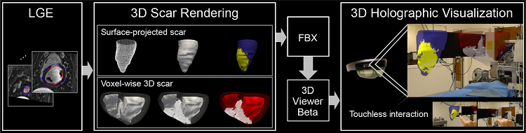
 A framework for holographic visualization of the 3D late gadolinium enhancement data. Source: Jang et al.
Superimposing computer-generated information on a user's view of the real world, offers a new platform to help physicians better visualize complex medical data, particularly before and during medical procedures. A new self-contained augmented reality (AR) device was designed by an international research team to provide an immersive AR experience in which surgeons can interactively explore data in 3D.
The researchers examined AR’s potential to help cardiologists visualize myocardial scarring in the heart as they perform ventricular tachycardia ablation or other electrophysiological interventions. By projecting 3D imagery onto a HoloLens face-worn glass screen, AR provides 3D depth perception and allows surgeons to interact with the medical data without physically touching a screen or computer mouse, maintaining a sterile environment and reducing the risk of infection.
The AR technology was tested in a pilot study as holographic 3D scars were generated in five animal models that underwent controlled infarction and electrophysiological study. An operator and mapping specialist viewed the holographic 3D scar during electrophysiological assessment and completed a perceived usefulness questionnaire. The system was deemed useful as the practitioner could interactively explore 3D myocardial scars.
Scientists from Beth Israel Deaconess Medical Center in Massachusetts, University of Pennsylvania and Technical University of Munich contributed to this research, which is published in PLOS ONE.
