Sponsored Content
Medical Devices and Healthcare IT
So long, trial and error
24 March 2020
If you have ever traveled in a modern jetliner, crossed a bridge or ridden in a car, you have entrusted your life to computer simulations. Such models are used to design everything from aircraft and bridges to automobiles and buildings, making sure they are as safe as possible. But when it comes to designing treatments for patients with serious and even life-threatening conditions, physicians often rely on an approach best described as “trial and error.”
 Figure 1: Natalia Trayanova. Source: Johns Hopkins UniversityMy research is dedicated to changing that. With my team, I am working to bring the precision of computer simulations to clinical practice so we might help physicians make the best possible treatment decisions for every patient. I believe that such models are poised to transform medicine, heralding the arrival of a new brand of personalized health care and dramatically improving patient outcomes.
In my lab, we are doing this through the creation of sophisticated 3D “virtual hearts” — models that enable cardiologists to test lifesaving interventions. Doctors can poke and prod these virtual organs in ways that are simply not possible with flesh-and-blood hearts. Clinicians not only minimize the invasiveness of diagnostic procedures but also — and most importantly — improve therapies.
One of our projects provides an example. Using MRI images, we have created detailed models of the hearts of patients who had survived heart attacks but were left with damaged tissue that predisposes their hearts to deadly arrhythmia. This is a condition in which the heart’s electrical signals run amok, causing irregular and abnormal rhythms that can result in sudden death.
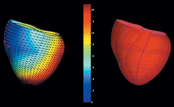 Figure 2: The virtual hearts. Source: Johns Hopkins UniversityCurrent treatment involves implanting a small device called a defibrillator that can sense the onset of an abnormal rhythm event and shock the heart back to its normal pace. This invasive procedure carries numerous risks, including infections and heart tissue damage, so it is important that only patients who need the device get it.
So how do doctors determine which patients actually need defibrillators? They use something called the “ejection fraction,” a measure of the amount of blood pumped out of the heart with each beat. If that number is less than 35%, then the patient is considered at high risk of sudden cardiac death by arrhythmia and warrants an implant.
The trouble is that ejection fraction is not a good predictor of the patient’s risk of dying suddenly, because it only considers blood flow and does not consider the heart’s electrical activity. Using only blood flow to predict who will experience a life-ending arrhythmia episode is like having an electrical problem in your house and calling a plumber to fix it. As a result, many patients get defibrillators that they do not really need. Worse yet, this inaccurate criterion means that many patients who are at risk of suffering arrhythmias do not get the devices they need.
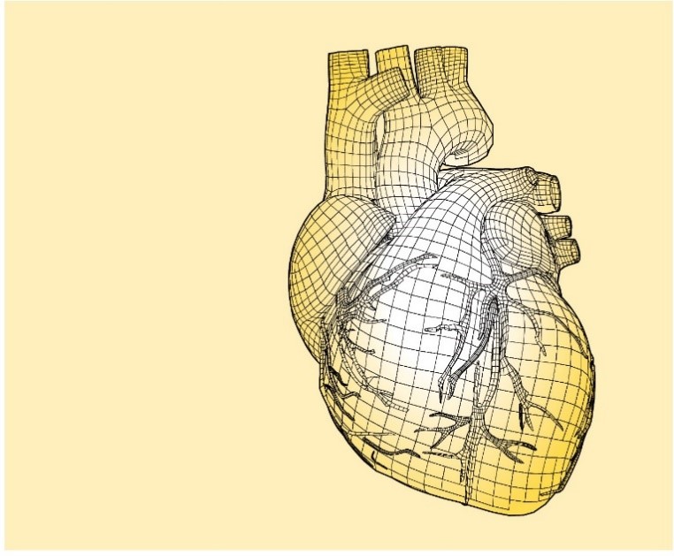 Figure 3: A virtual heart model. Source: Johns Hopkins UniversityOur personalized virtual hearts could help doctors make better decisions in these cases. These digital replicas allow us to factor in not only the unique geometry of each patient’s individual heart, but also the impact of the heart-attack-related scar tissue and the pattern of electrical waves as they move throughout the organ.
We have demonstrated that this method, called virtual-heart arrhythmia predictor (VARP), significantly outperforms any other current approaches for predicting which patients are at risk of arrhythmias. It has the potential to save lives, and to save patients from getting defibrillators they do not need.
We look forward to a time when all heart patients, from babies to senior citizens, have virtual hearts, which doctors can then use to plan their personalized treatment. The pursuit of this merger of computational simulation and clinical medicine is the future of medicine.
About the author
Natalia Trayanova, the inaugural Murray B. Sachs Professor in the Department of Biomedical Engineering at the Johns Hopkins University, is a fellow of the Heart Rhythm Society, American Heart Association, and the American Institute for Medical and Biological Engineering. She is a pioneer in the development of personalized image-based computer models of the heart and was a 2019 inductee into the Women in Technology International Hall of Fame. She currently holds faculty appointments within the Johns Hopkins University Whiting School of Engineering and its School of Medicine, and is a core faculty member of the university's Institute for Computational Medicine. For more information visit Engineering.JHU.EDU.
Powered by CR4, the Engineering Community
Discussion – 0 comments
By posting a comment you confirm that you have read and accept our Posting Rules and Terms of Use.
Advertisement
Advertisement
Popular News
Find Free Electronics Datasheets

Advertisement